Dental-OPG-3D-X-Ray
BOOK AN APPOINTMENT
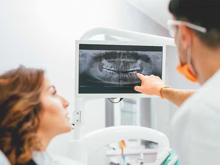
A dental OPG 3D X-ray, also known as an Orthopantomogram, is a specialized imaging technique used in dentistry to capture a panoramic and three-dimensional image of a patient’s oral structures. It provides a comprehensive view of the teeth, jaws, temporomandibular joints (TMJ), and surrounding tissues.
Key features and principles of a dental OPG 3D X-ray include:
Panoramic Imaging: The OPG 3D X-ray captures a wide-angle panoramic image of the entire dental arch, including both upper and lower jaws. This allows dentists to visualize all the teeth, their positioning, and their relationship to adjacent structures.
Three-Dimensional Imaging: In addition to the panoramic view, the OPG 3D X-ray also produces a three-dimensional representation of the oral structures. This provides enhanced visualization and helps dentists accurately assess complex anatomical relationships and plan for surgical interventions or treatments.
Diagnostic Capabilities: The OPG 3D X-ray aids in diagnosing various dental conditions, such as impacted teeth, dental infections, cysts, tumors, bone abnormalities, and temporomandibular joint disorders. The detailed images enable dentists to identify and analyze dental and skeletal irregularities that may not be visible in traditional two-dimensional X-rays.
Treatment Planning: The detailed and comprehensive images obtained from the OPG 3D X-ray assist dentists in developing precise treatment plans. It helps them evaluate the need for orthodontic interventions, dental implant placement, extractions, and other dental procedures. Dentists can also assess the bone quality and quantity before implant surgeries.
Radiation Safety: The OPG 3D X-ray utilizes modern imaging technology that minimizes radiation exposure to the patient. It employs a focused beam of X-rays and digital sensors to capture the images efficiently while ensuring patient safety.
Patient Comfort and Convenience: The OPG 3D X-ray is a non-invasive procedure that requires minimal patient cooperation. Patients stand or sit in a designated position while the imaging equipment rotates around their head, capturing images in a matter of seconds. This enhances patient comfort and allows for a quick and efficient examination.
Collaboration with Dental Specialists: OPG 3D X-rays are an essential tool for communication and collaboration between dentists and other dental specialists, such as orthodontists, oral and maxillofacial surgeons, and prosthodontists. The detailed images facilitate comprehensive treatment planning and interdisciplinary coordination when addressing complex dental cases.
Dental OPG 3D X-rays are commonly used in general dental practices, oral surgery clinics, orthodontic centers, and dental imaging centers. They provide valuable diagnostic information, aid in treatment planning, and contribute to delivering precise and effective dental care.